Going Beyond Limits
New lens lets microscopes peer at much smaller objects.
The optical microscope is a workhorse of biology, but it has its limits—specifically the diffraction limit, which says it can't resolve anything smaller than about half the wavelength of visible light. Now one Lincoln Laboratory researcher has developed a lens array that he says can increase the resolution of tiny objects by 400 percent, enough to study the innards of bacteria.
Such enhanced vision can allow people to quickly identify dangerous organisms, such as anthrax, by sight, instead of running cultures and waiting for the results. "You can see a lot more details, and you can better identify them," says Zong-Long Liau, a materials scientist and device physicist in the Laboratory's Electro-Optical Materials and Devices Group. "It will allow us to measure a biological sample in more like a three-dimensional way."
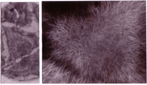
This comparison of micrographs of a cell shows the higher resolution made possible by the solid immersion lens (right) over conventional microscopy (left). Both images were made using identical microscope objectives and magnifications. The cell is about 40 micrometers across. |
Liau has created tiny lenses that sit between the microscope objective and the object being studied. The lenses are made of gallium phosphide, which has a very high index of refraction of 3.5. If he places the sample directly against the lens at the microscope's focal point, that tiny area is magnified by a factor of the refractive index, in this case 3.5. As a result, the resolution of the image is now 350 percent of what it would be in air. Because the light is traveling through a high-index material, it slows down somewhat, essentially behaving as if it has a shorter wavelength and thus a smaller diffraction limit. Say you're looking at green light, which at roughly 550 nm is about in the middle of the visible spectrum. Its normal diffraction limit would be about 275 nm, but the gallium phosphide reduces that to about 80 nm—an improvement that makes smaller objects visible. Liau uses gallium phosphide because it is transparent at most wavelengths of visible light, whereas other high-index materials are transparent only in the infrared. Aside from the fact that optical microscopes rely on visible light, the longer wavelength infrared light would not achieve the same resolution, even when the wavelength is reduced to half its original value.
The concept is not entirely new. Scientists have long known that placing a tiny drop of oil between the microscope and the sample could increase resolution by 50 percent because of the oil's 1.5 index of refraction. In fact, it's by analogy with the oil drop method that this is called a "solid immersion lens." Though the sample is not truly immersed in the material, as it would be in the oil, it has to be so pressed so close against the lens that it is as good as inside it.
Such microlenses have been considered before, but implemented in a reversed way. In the 1990s, Liau says, some researchers considered using such lenses to focus a laser spot down to a very small dimension; their goal was to increase the density of the markings that store data on a compact disc. That didn't work out very well, Liau says, because no one developed an easy way to manufacture the tiny lenses in quantity. Most microlenses are stamped out of low-index polymers, a process that is relatively simple. But these high-index semiconductor lenses have to be smooth regular hemispheres that measure only about 200 micrometers in diameter; making them, Liau says, "is not entirely trivial."
James Leger, a professor of electrical and computer engineering at the University of Minnesota who worked with Liau at Lincoln Laboratory in the early 90s, agrees that the fabrication process can be tough, especially if the maker wants to introduce any variation to the shapes. "If you want to make aspheric shapes or you want to make a bunch of hemispheres in a row, that's a lot harder to do," he says.
Liau's lab manufactures the lenses using photolithography and ion chemical etching, common procedures used for making computer microchips. But in chip making, the structures are flat. The lenses need to be hemispheric. One way to make spheres out of little bits of semiconductor material is to tumble them together, so that impact and friction smooth out their surfaces. But Liau says this process can create cracks within the material, rendering it useless as a lens. Moreover, he's not convinced that the grinding process produces a perfectly spherical shape.
Instead, Liau relies on a process he invented, and which the Laboratory patented. He uses photolithography and etching to carve gallium phosphide into an array of lens shapes, each very close to the final hemisphere shape he wants to end up with. Then he heats the array to 1050 degrees Celsius. That's not quite the melting point for the gallium phosphide, but it's enough to allow atoms on the surface to move around easily. Since a smooth surface represents a lower-energy state than a rough surface, the mobility lets the atoms rearrange themselves so the lens is smooth. "It sounds simple, but there is a lot of materials science, even physics, involved," Liau says. His plan is to put an array of thousands on a 1-centimeter chip. Such a chip could be used in a manner similar to a microscope slide, with a sample pressed against the backside of multiple lenses.
Leger says the processes Liau has developed might help the solid-immersion lens find wider use. "He has pioneered a lot of the fabrication techniques for some of these binary compounds," Leger says, referring to materials such as gallium phosphide that contain two elements.
One area of continued research will be on ways to speed up production, which right now requires very slow etching and uses special equipment built in Liau's lab. To commercialize the lenses, Liau will need to improve the throughput of the process.
Liau has come up with a further improvement to the lenses that increases resolution even more—to 400 percent better than in air. The key here is to alter the lenses' shape somewhat, in ways he doesn't want to describe until after he has filed for patent protection. To show how effective this is, Liau displays a pair of photographs of a cultured cell taken through a microscope. In the first, which used a conventional lens, the cell is a small triangular shape without much internal structure visible. In the second, it becomes a thick array of branches, the cytoskeleton that makes up the cell's structure. Organelles that perform functions inside the organism are visible.
There isn't much contrast between different structures within a cell, and even with Liau's lenses, the sample has to be stained to make the structures visible. But the contrast is improved enough by his lenses that he can see structures that wouldn't be visible through a conventional microscope lens without the use of complex fluorescence techniques.
Having demonstrated that the lenses work, Liau wants to start using them to see what he can learn about different bacteria, to show that valuable information can be gleaned. He also plans to apply to the Defense Advanced Research Projects Agency or the National Institutes of Health for funding for the research. It could take another two or three years to commercialize the lenses, he predicts. "This is a very strong lens, the strongest people ever produced," Liau says. "This is the first time that people have ever looked at things this way."
|